PoC & Feasibility Studies
Generating in-vivo evidence on implant function, immune response and early device comparisons, in time to inform design decisions before design freeze.
Focused prototype studies and expanded feasibility evaluations.

BERLINANALYTIX
The histopathology CRO for
biomaterials & medical devices.
Specialised processing. Histological analysis.
REAL HISTOLOGY. A UNIVERSE OF QUESTIONS.
01 / STUDIES FOR YOUR PRODUCT
Specialist histology for bone, collagen and bone substitute materials. Characterising foreign-body responses and implant integration in hard and soft tissue—with clear interpretation of what the findings mean for your device.
Choose the study that fits your question.
Services in focus
An overview of our in-vivo, processing and histopathology services.
60 seconds · Silent film
Generating in-vivo evidence on implant function, immune response and early device comparisons, in time to inform design decisions before design freeze.
Focused prototype studies and expanded feasibility evaluations.
Building a stronger body of preclinical evidence through larger-scale studies of implant behaviour across multiple timepoints and against predicate or reference devices.
Supporting scientific validation, FDA submissions and EU MDR technical documentation.
Characterising residual cellular and material components in biomaterials and evaluating the effects of processing changes.
Bone substitute materials, collagen patches and processing comparisons.
Generating preclinical evidence for regulatory submissions through GLP study conduct, quality assurance and traceable reporting.
Submission studies with full GLP documentation and oversight.
Generating histopathological evidence from clinical biopsies and explants for efficacy evaluation, clinical surveillance and post-market clinical follow-up.
Tissue response, implant integration and degradation in clinical specimens.
02 / FROM TISSUE TO EVIDENCE
Understanding how your device or biomaterial interacts with tissue, changes over time and compares with a reference device.
PUBLISHED PRECLINICAL EVIDENCE
Comparing two bone substitutes at 2 and 16 weeks, with tissue-level observations and measured bone formation.
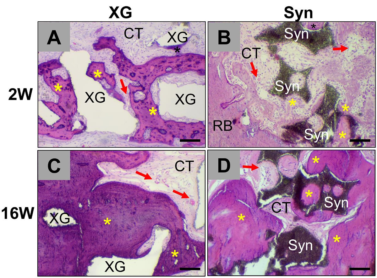
A/C: xenogenic bone substitute material (XG). B/D: synthetic bone substitute material (Syn). Top: 2 weeks; bottom: 16 weeks. Asterisks: new bone; red arrows: blood vessels; CT: connective tissue. Scale bars: 100 µm.
Figure 1 reproduced unchanged from Bielenstein et al. (2022), with Mike Barbeck as corresponding author. © The authors, CC BY 4.0. Bone-area values: Table 3.
| Material | New bone |
|---|---|
| Xenogenic bone substitute material | 31.98 ± 24.83% |
| Synthetic bone substitute material | 24.10 ± 9.61% |
Rat calvarial model · 10 defect sites per material and timepoint. Absence of a significant difference does not establish clinical equivalence.
Technovit 9100 plastic embedding, H&E staining and histomorphometry. Additional CD163 and CD11c immunohistochemistry characterised selected aspects of the tissue response.
03 / THE EXPERTISE BEHIND THE EVIDENCE
Study design, specialised preparation and scientific interpretation, connected around what you need to learn.
Explore the processingCinematic process imagery meets authentic histology.
Specialised processing · Histological analysis
Mineralised bone. Implant interfaces. Soft-tissue architecture. Preparation built around the features your investigation needs to assess.
01 / Undecalcified bone & implants
Specialised resin embedding and sectioning for bone, titanium and ceramic implants. Preserving mineralised architecture and the relationship between the implant and surrounding tissue.
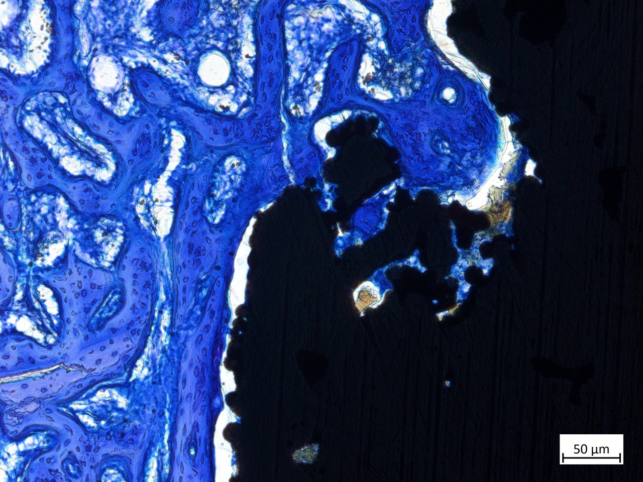
Undecalcified resin preparation retains the mineralised features under investigation. Cutting and grinding enable examination of implant-containing sections. The specimen, section orientation and study endpoints determine the preparation and subsequent histomorphometry.
02 / Soft tissue & paraffin

Paraffin processing for the tissue architecture surrounding soft-tissue implants. Assessing regeneration, matrix organisation, fibrosis and encapsulation in regenerative, cosmetic and cardiovascular applications.
The evidence: the extent and distribution of the tissue response, compared across device groups and follow-up timepoints.
Preparation and section orientation follow the specimen and the structures being evaluated. Where mineralised tissue is present, any decalcification step is selected against the study endpoints. Soft-tissue processing does not itself require decalcification.
03 / Staining & immunohistochemistry
A broad histochemical and immunohistochemical repertoire, scoped to the structures and cell markers under investigation.
How does tissue composition
change across timepoints?
The evidence: tissue composition and organisation linked to the study's regeneration, integration or response endpoints.
Routine morphology, specialised histochemistry and IHC are combined where they answer the study question. Stain and marker selection, tissue preparation, controls and the analysis plan are defined together. Comparisons use the study's actual groups and timepoints.
04 / ONE COORDINATED PATHWAY
A defined scientific contribution at every stage,
with study scope and responsibilities agreed
from the beginning.
Focused non-GLP investigations through our integrated partner site. GLP in-vivo studies through our trusted partner network.
Specialised preparation in Berlin for hard and soft tissue, including undecalcified bone and specimens containing implants.
Histopathology, selected quantitative readouts and scientific reporting that connect observations to your study question.
Quality requirements belong to the commissioned study.
Explore the certification and accreditation scope of our Berlin laboratory.
05 / YOUR SCIENTIFIC PARTNERS
Connect your product question with the scientists responsible for the investigation.
Meet the full team Explore our published science
Founder & scientific leadership
Biomaterials. Tissue response.
Laboratory & study leadership
Study design. Laboratory execution.THE NEXT QUESTION IS YOURS.
Tell us about your device, your development stage
and the decision you want the evidence to inform.