Biopsies and explants
Investigating available clinical tissue, explants and retrieved devices with preparation and evaluation matched to the specimen.
Histopathological evidence from the tissue available for assessment.

05 / Clinical Studies
Generating histopathological evidence from clinical biopsies and explants for efficacy evaluation, clinical surveillance and post-market clinical follow-up.
Tissue response, implant integration and degradation in clinical specimens.
The study scope
Connecting biopsy and explant findings with the clinical question and the tissue endpoints of the commissioned investigation.
Investigating available clinical tissue, explants and retrieved devices with preparation and evaluation matched to the specimen.
Histopathological evidence from the tissue available for assessment.
Assessing the specified tissue features in support of efficacy evaluation, clinical surveillance or post-market clinical follow-up.
Findings interpreted within the clinical and specimen context.
The service covers specimen processing and histopathological analysis within the clinical study or follow-up programme.
Published clinical evidence
Quantifying tissue regeneration, augmentation and integration, and connecting those findings with clinical outcomes.
01 / Bone regeneration · Randomized clinical study
40 patients · 6 months · 29 evaluable biopsies
Comparing two sinus-augmentation approaches using the same xenogenic bone substitute: bony-wall repositioning and collagen-membrane coverage.
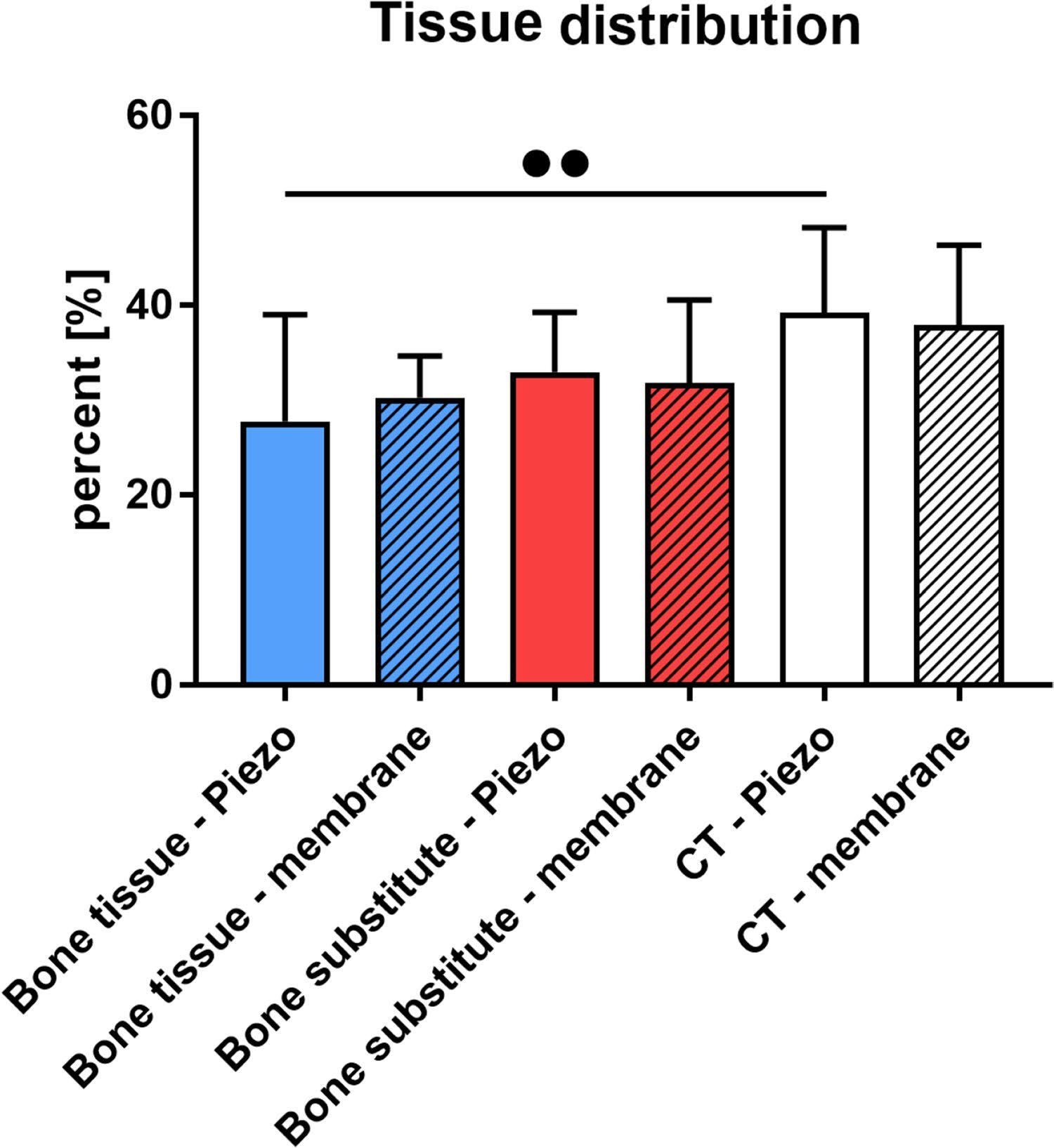
| Tissue | Bony wall 13 biopsies | Membrane 16 biopsies |
|---|---|---|
| New bone | 27.8 ± 11.2 | 30.3 ± 4.5 |
| Remaining substitute | 32.9 ± 6.3 | 31.8 ± 8.8 |
| Connective tissue | 39.2 ± 9.0 | 37.9 ± 8.5 |
Forty patients were randomized, 20 per group. Twenty-nine biopsies were evaluable; eleven were excluded because of insufficient measurable material or processing difficulties. Histomorphometry was a secondary endpoint. The study compared surgical approaches, not different bone substitutes. Mike Barbeck is credited with histomorphometric analysis. These findings do not establish long-term implant equivalence.
02 / Soft-tissue augmentation · Clinical case series
20 patients · 2 months · Collagen matrix augmentation
Clinical thickness measurements paired with biopsy analysis of graft integration.
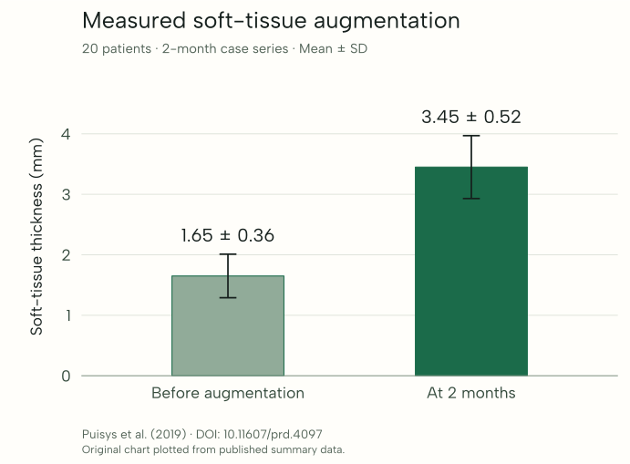
Short-term tissue gain and integration were evaluated; improved implant survival was not established by this case series.
| Assessment | Thickness (mm) |
|---|---|
| Before augmentation | 1.65 ± 0.36 |
| At two months | 3.45 ± 0.52 |
Twenty patients received a porcine-derived collagen matrix during implant treatment. Clinical measurements and biopsies were evaluated at two months. The plotted values are reported group means and standard deviations; individual patient data are not reconstructed.
Further reading
Co-authored by Prof. Dr. Mike Barbeck. Titles below summarise each paper; links open the published articles.
Molnár et al. · Clinical Oral Investigations · Randomized clinical and histological study; 40 patients, six months.
Puisys et al. · International Journal of Periodontics & Restorative Dentistry · Clinical and histological case series; 20 patients, two months.
Lorenz et al. · Clinical Oral Investigations · Prospective clinical and histological study; eight patients, six months.
Lorenz et al. · Annals of Maxillofacial Surgery · Randomized split-mouth clinical study; eight patients, six months.
Optional technical detail
Retaining the structures that matter in the available specimen and connecting the analytical findings with the clinical question.
Explore processing & analysisSelecting undecalcified processing when mineralised structures or implant interfaces require preservation, and appropriate soft-tissue processing for biopsies and other tissue specimens. Preparation is planned around the specimen received and the study endpoints.
Selecting stains and, where relevant, immunohistochemistry for the tissue and cellular features under investigation. Interpreting the section findings in the context of the submitted clinical information.
Applying the qualitative and quantitative evaluations appropriate to the commissioned endpoints and available tissue. Reporting observations with the specimen and methodological context needed for their interpretation.
Your next investigation
Connect the clinical question with the tissue available and the analysis needed for your study or follow-up programme.
Discuss clinical specimens